
Prototipo de un dispositivo para la investigación de los efectos del ultrasonido sobre procesos microbiológicos
Main Article Content
Keywords
Biofísica del ultrasonido, micro-incubadora, nefelometría láser, procesos microbiológicos, sonicación, ultrasonido continuo, ultrasonido pulsado.
Resumen
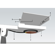
Este artículo describe el desarrollo de un prototipo multifuncional que suministra dosis de ultrasonido a una muestra contenida en una caja de Petri, en frecuencias comprendidas entre 18 y 30 KHz y con intensidades que van desde los 0,3 mW/m2 hasta los 1,5 mW/m2. Este dispositivo garantiza la supervivencia de la muestra mediante un sistema de micro-incubación, operado por un sistema de control y monitoreo de temperatura. Además, el prototipo puede detectar cambios, inherentes al proceso microbiológico estudiado, por medio de un sistema basado en nefelometría láser. El usuario puede escoger, para un experimento específico, el valor de los parámetros mencionados y analizar los datos generados a través de una aplicación computacional especialmente diseñada para presentarlos en tablas de datos, imágenes y estadísticas.
MSC: 92-XX
PACS: *43.35.-c; 43.35.+d; *43.80.-n; 43.80.+p; 87.50.Y
Descargas
Referencias
[2] V. Frenkel, Therapeutic ultrasound: Mechanisms to applications. New York: Nova Science Publishers, 2011.
[3] N. Doan, P. Rehem, S. Meghji, and M. Harris, “In vitro effects of therapeutic ultrasound on cell proliferation, protein synthesis, and cytokine production by human fibroblasts, osteoblasts, and monocytes,” J Oral Maxillofac Surg, vol. 57, pp. 409–419, 1999.
[4] L. J. M. Juffermans, P. A. Dijkmans, R. J. P. Musters, C. A. Visser, and O. Kamp, “Transient permeabilization of cell membranes by ultrasound-exposed microbubbles is related to formation of hydrogen peroxide,” American Journal of Physiology - Heart and Circulatory Physiology, vol. 291, no. 4, pp. H1595–H1601, 2006. [Online]. Available: http://ajpheart.physiology.org/content/291/4/H1595
[5] W. Wei, B. Zheng-zhong, W. Yong-jie, Z. Qing-wu, and M. Yalin, “Bioeffects of low-frequency ultrasonic gene delivery and safety on cell membrane permeability control,” Journal of Ultrasound in Medicine, vol. 23, no. 12, pp. 1569–1582, 2004. [Online]. Available: http://www.jultrasoundmed.org/content/23/12/1569.abstract
[6] M. Madigan, J. Martinko, J. Parker, and T. Brock, Biología de los microorganismos. Madrid: Pearson, Prentice Hall, 2004.
[7] WHO, Laboratory safety manual. Geneva: WHO library, 2004.
[8] I. E. Commission, 61010-1 Safety Requirements for Electrical Equipment for Measurement, Control and Laboratory Use-Part 1: General Requirements, Geneva, 2010.
[9] E. Corporation, XR-2206 Monolithic Function Generator Application Note, Kato Road, Fremont, CA, 1997.
[10] K. G. Baker, V. J. Robertson, and F. A. Duck, “A review of therapeutic ultrasound: Biophysical effects,” Physical Therapy, vol. 81, no. 7, pp. 1351–1358, 2001. [Online]. Available: http://ptjournal.apta.org/content/81/7/1351.abstract
[11] S. Robinson, Driving Piezoelectric actuators, Tuczon, Ariz, 2006.
[12] W. L. Nyborg, “Biological effects of ultrasound: Development of safety guidelines. part ii: General review,” Ultrasound in Medicine and Biology, vol. 27, no. 3, pp. 301–333, 2001, cited By (since 1996):112. [Online]. Available: www.scopus.com
[13] L. A. Love and F. W. Kremkau, “Intracellular temperature distribution produced by ultrasound,” The Journal of the Acoustical Society of America, vol. 67, no. 3, pp. 1045–1050, 1980. [Online]. Available: http://link.aip.org/link/?JAS/67/1045/1
[14] WHO, Environmental Health Criteria. Geneva: WHO library, 1982.
[15] K. J. Astrom, “Contro system design lecture notes for me 155a,” pp. 217–237, 2002.
[16] S. Angersbach, B. Hipler, S. Brand, and C. Ruckert, “Monitoring of microbial growth curves by laser nephelometry,” GOR-TOKYO-, vol. 7, no. 4, p. 35, 2005.
[17] M. Sadar, Introduction to laser nephelometry: an alternative to conventional particulate analysis methods, Loveland, CO, 1999-2005.
[18] B. H. Brown, R. H. Smallwood, D. C. Barber, P. V. Lawford, and D. R. Hose, “Medical physics and biomedical engineering,” Measurement Science and Technology, vol. 12, no. 10, p. 1744, 2001. [Online]. Available: http://stacks.iop.org/0957-0233/12/i=10/a=703
Article Sidebar
Article Details
Jorge Andrés López-Castaño, Universidad Manuela Beltrán
Ingeniero Biomédico Universidad Manuela Beltrán, ColombiaYuri K Murcia, Universidad Manuela Beltrán
Ingeniera Biomédica UMBDaniel Díaz, Universidad Manuela Beltrán
Ingeniero Físico, Msc en Física y Tecnología de los Láseres, Docente Investigador, Universidad Manuela Beltrán.Los autores que publican en esta revista están de acuerdo con los siguientes términos:
- Los autores conservan los derechos de autor y garantizan a la revista el derecho de ser la primera publicación del trabajo al igual que licenciado bajo una Creative Commons Attribution License que permite a otros compartir el trabajo con un reconocimiento de la autoría del trabajo y la publicación inicial en esta revista.
- Los autores pueden establecer por separado acuerdos adicionales para la distribución no exclusiva de la versión de la obra publicada en la revista (por ejemplo, situarlo en un repositorio institucional o publicarlo en un libro), con un reconocimiento de su publicación inicial en esta revista.
- Se permite y se anima a los autores a difundir sus trabajos electrónicamente (por ejemplo, en repositorios institucionales o en su propio sitio web) antes y durante el proceso de envío, ya que puede dar lugar a intercambios productivos, así como a una citación más temprana y mayor de los trabajos publicados (Véase The Effect of Open Access) (en inglés).